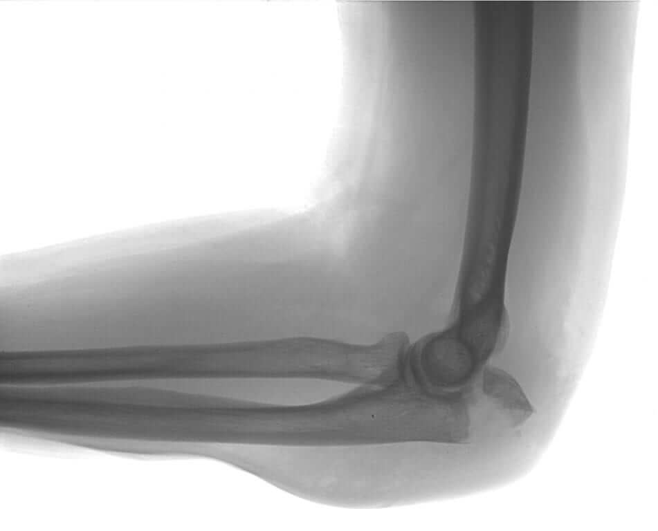
Elbow & Radial Head Fractures
At Glenelg Orthopaedics we commonly encounter radial head fractures. The forearm is made up of two bones; the radius and the ulna. The radial head is located at the superior end of the radius. These bones articulate with the lower aspect of the humerus to form the elbow joint.
Radial head fractures often occur during a fall. The force of the fall is commonly transmitted through the forearm via an outstretched arm, consequently impacting the radial head as it articulates with the distal humerus. With this, the supporting interosseous ligament can also be damaged. Pain will often be felt in the forearm and elbow. In some circumstances, damage to the distal radioulnar joint can occur, resulting in wrist discomfort.

There are three grades of radial head fractures.
- Grade I: displaced.
- Grade II: minimally displaced.
- Grade III: significantly displaced.
How are radial head fractures treated?
Fortunately, the majority of radial head fractures are either Grade I or Grade II and do not require surgical intervention. A sling can be used to minimise discomfort. However, it is of vital importance that elbow motion is encouraged as soon as possible. Movements involving flexion and extension, as well as pronation and supination are recommended.
If the arm is encouraged to move would that make the fracture move and possibly result in surgery?
It is extremely unlikely for the fracture to move. However, it is common for the elbow to get stiff after such an injury. Lack of motion or leaving the elbow immobile can increase the probability of a fracture moving. If this occurs, it is quite difficult to treat. For this reason, it is actually harmful to leave the elbow stationary.
What is done if the fracture is displaced?
The method of treatment depends on a number of factors, including the patient’s age, the degree of displacement, and the number of fragments. Options for treatment include the use of screws or a metal plate or, in some circumstances, the use of an artificial radial head.
What other fractures occur around the elbow?
The tip of the elbow (olecranon) is another common fracture. This occurs in a similar fashion to the radial head fracture but often is associated with a muscle contraction, where the broken fragment is attached to the triceps muscle (the muscle in the back of the arm). When this fracture occurs, the triceps muscle pulls the bone away, resulting in displacement. The vast majority of these fractures will require fixation as fragments, if not yet displaced, will eventually displace by the pull of the triceps muscle. Fixation can be done through tension band wiring, or with metal plates. Under these circumstances, the arm will be placed in a splint to protect it for a period of time before motion is commenced. This time period will depend on the stability of the fracture.
Are there any other types of fractures?
Yes. Other fractures include fractures to the distal humerus, or combination fractures involving the radial head and the olecranon. If combination injuries occur in conjunction with ligament damage, it is known as a terrible triad. Commonly, these injuries will require a brace.
What is a terrible triad?
A terrible triad is a complex dislocation of the elbow where ligaments on the outside, or commonly on the inside of the elbow, are torn. This leads to instability along with associated breaks to the radial head and the olecranon.
Quite often a fractured olecranon will occur in conjunction with damage to the coronoid process, the anatomical function of which is to create stability in the arm.
Unfortunately, due to the nature of this injury, treatment can be associated with significant amounts of pain. However, it is important that motion is encouraged to prevent stiffness. In the long term, this will allow the elbow to recover to its full potential.
For best result, it is important to consult a surgeon who has experience with such injuries.
Are there any risks associated with the treatment of these injuries?
Unfortunately, all treatments will have associated risks.
Firstly, treatment involves the bone healing naturally, the rate of which can vary depending on the individual. Factors such as smoking can hinder the rate of bone healing, increasing the risk of wound breakdown, and infection. Furthermore, smoking can inflame the nerves, ultimately causing more pain.
Secondly, as outlined above, the development of stiffness can impede management. It is of vital importance that full motion is encouraged despite the pain that may be experienced. The longer one delays movement, the more likely stiffness will occur. At Glenelg Orthopaedics we relate this to cement setting; while cement is still soft, it is easier to mould. However, once it starts to harden, the dye is often cast as the shape of the cement.
Thirdly, there are important nerves around the elbow that are responsible for innervating the muscles in the hand and providing sensation. Unfortunately, with surgical intervention comes the risk of nerve damage. If this were to occur, the patient could lose function of these muscles, or experience numbness in associated regions. This risk is rare but needs to be taken into consideration when approaching management options.
Fourthly, the time of recovery is not as quick as people hope due to many of the factors outlined above. Under normal circumstances, we would expect the patient to have reasonable function at three months, however, it is unlikely full function will return until about twelve months.
Finally, one of the most significant issues that arise with upper limb fractures is the frustration associated with management. Until full function has returned, the patient must be cautious with their injury. Often, the patient may not be fit to drive a motor vehicle due to the risk of losing control of the vehicle.
At Glenelg Orthopaedics we have examined and treated a significant number of elbow fractures. We aim to provide Quality Orthopaedic Care.
